|
In situ hybridization (ISH) and Fluorescence in situ hybridization (FISH)
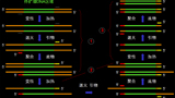
ISH is broadly used in detecting DNA or RNA in pathologic tissues, its visualized methods frequently used today are nonradioactive or fluorochrome labeling the probe (FISH). ISH or FISH has its advantages in that on one side we may observe the gene changes occurring in tissues directly, such as HPV in cervical disease, EBV in lymphoma and nasopharyngeal carcinoma; on the other side we may detect a gene amplification in a tumor such as CerbB-2 in breast carcinoma and gastric adenocarcinoma, EGFR in non-small cell lung carcinoma, MDM2 in dedifferentiate liposarcoma. A standardized laboratory and process for ISH or FISH should be introduced to guarantee a highly duplication and reliability of the results.
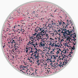
ISH present EBER positivity in a peripheral T cell lymphoma |
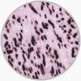
ISH shows EBER positivity in a EB associated leomyosarcoma |
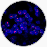
FISH shows Her2 amplification (red) in a breast invasive ductular carcinoma |
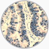
CISH shows Her2 amplification in a gastric adenocarcinoma |
Polymerase chain reaction (PCR) Technique
PCR technique is greatly attractive in surgical pathology in that it can be utilized to analyze DNA and RNA from samples of formalin-fixed, paraffin-embedded materials. Up to now, the PCR in our Department is used for various aims such as for detecting Immunoglobulin (Ig) or T cell receptor (TCR) gene rearrangements to determine the clonality of B- or T cell proliferations, for exposing chromosomal translocations occurred in hematologic malignancies (e.g., t(14;18) in follicular lymphoma); for discerning the point mutations in oncogenes and tumor-suppressor genes (e.g., activated RAS oncogenes in pancreatic carcinomas and EGFR mutation in non-small cell lung carcinoma); for identifying gene amplification (e.g., MYCN in neuroblastoma and HER2 in breast carcinoma); for showing microsatellite instability in gastrointestinal tumor and gene hypermethylation in various tumors.
A principle of PCR technique |
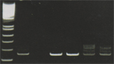
T cell receiptorgene rearrangement from a peripheral T cell lymphoma |
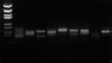
Immunoglobulin heavy chain variable gene (Fr3) rearrangement in a chronic lymphatic leikemia |
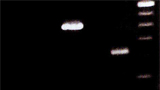
EWS-FL1 gene rearrangement in a Ewings in a synovial sarcoma |
|